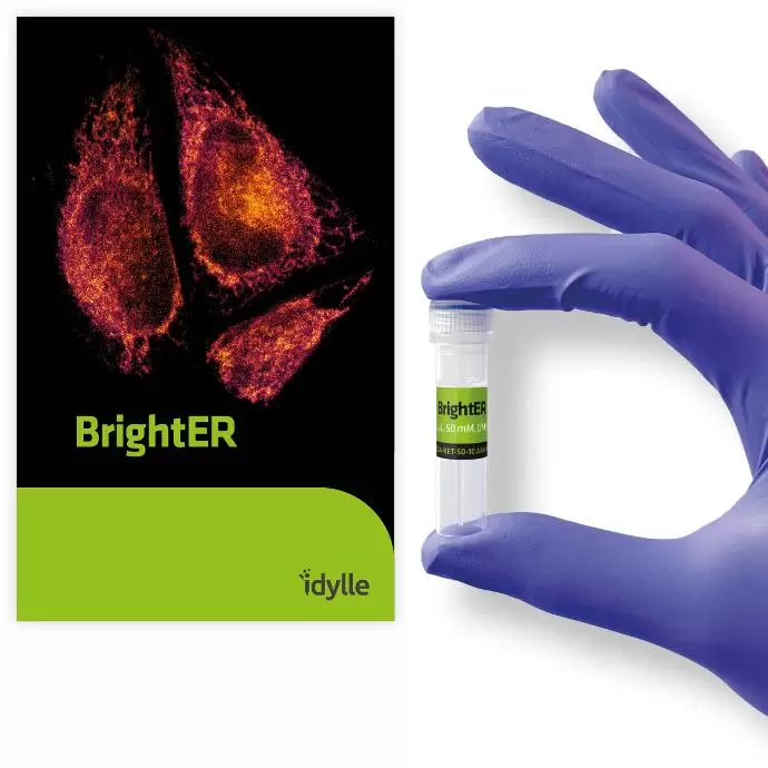
BrightER Endoplasmic Reticulum Probe
For Live Imaging & Flow Cytometry
Product Description
BrightER is a selective, cell-permeable endoplasmic reticulum probe conjugated to a bright photostable rhodamine dye. Simply add it to your culture medium and get a bright, stable ER fluorescence for at least 3 hours without altering ER physiology. One BrightER kit is sufficient to prepare 10 mL of final imaging medium when used at the recommended 50 µM concentration.
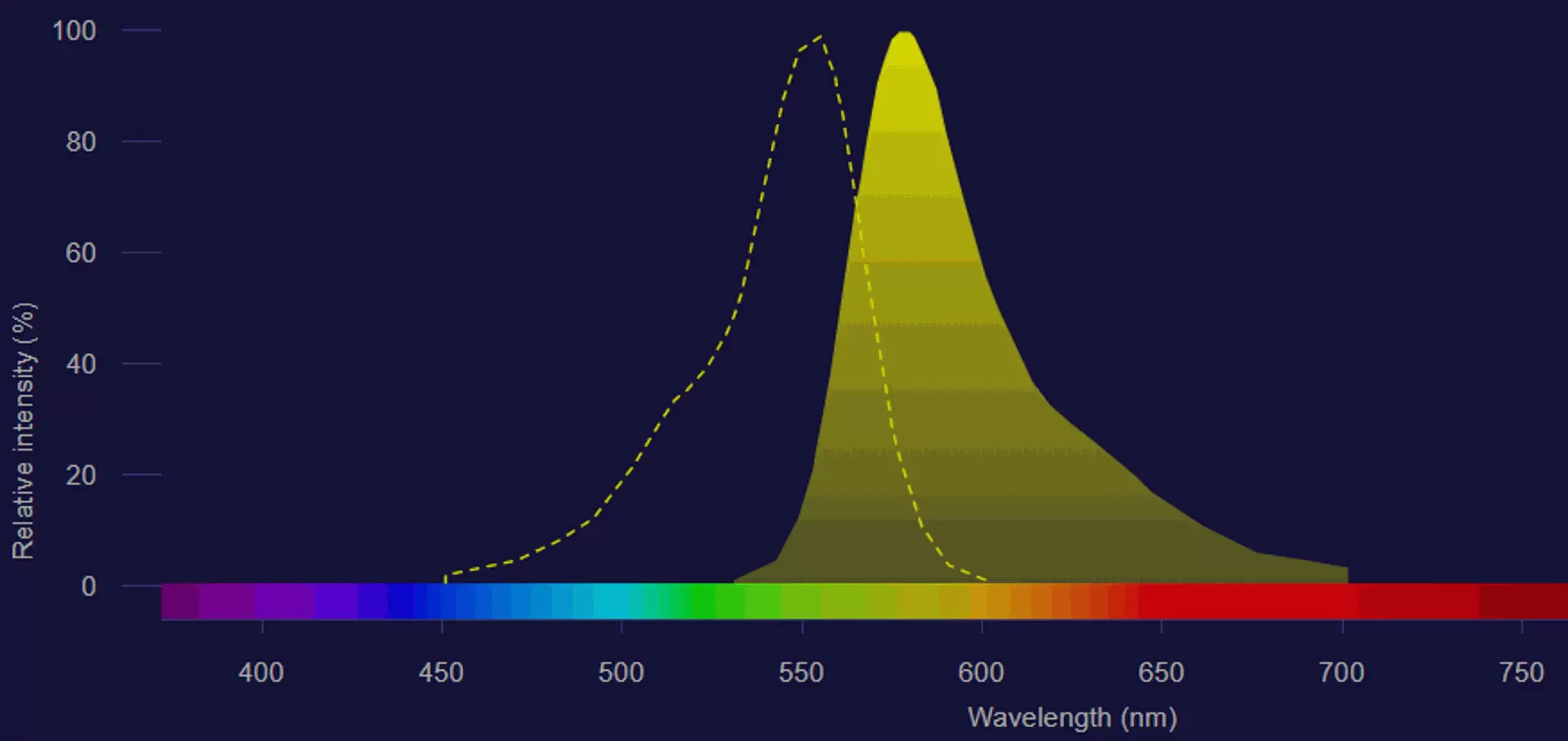
Fig.1: Absorption (dotted line) and emission (solid line) spectrum of BrightER
Image obtained on Fluorescence SpectraViewer (Thermo Fisher Scientific)
Applications
- Live imaging: ER morphological features and dynamic processes (membrane contact sites, intracellular trafficking, secretory pathways, viral infections, translation, protein folding and unfolded protein response (UPR), nuclear envelop formation upon mitosis, cellular calcium regulation and many others).
- Flow cytometry: quantification assays (ER stress detection, ER mass, co-localization studies)
Tested and validated on (Cell Types)
Patient-derived glioblastoma cells, HEK293, HeLa, U-2 OS (human osteosarcoma), SVG-A (human astrocytes), SUM159 (human breast cancer cells), B16F10 (murine melanoma), LLC (Lewis lung carcinoma) & Vero E6 (monkey kidney epithelial cells).
Suggestions for Use
The BrightER dye may be added directly in full media. In most cases, a final 50µM concentration is sufficient for immediate and bright ER staining; however, optimization may be needed for some cell types, conditions, and applications. BrightER is detected through standard TRITC and DsRed filters.
Specifications
Colour: Red
Detection method: Fluorescent
Wavelength range: Ex:557/Em:576
Dye type: Tetramethylrhodamine
Assays: confocal imaging, flow cytometry
Form: Liquid
SubCellular localization: Endoplasmic reticulum
Lifetime: up to 4 months at -20°C
Fixable: no
Kit Content
One vial of 100µL BrightER 5mM in DMSO+PEG solution.
Supporting Data
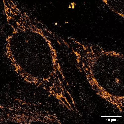
Fig.2: BrightER staining in live HeLa cells (60X objective)
Image credit: Raphaël Gaudin & Yonis Bare – IRIM CNRS, Université de Montpellier - 2023
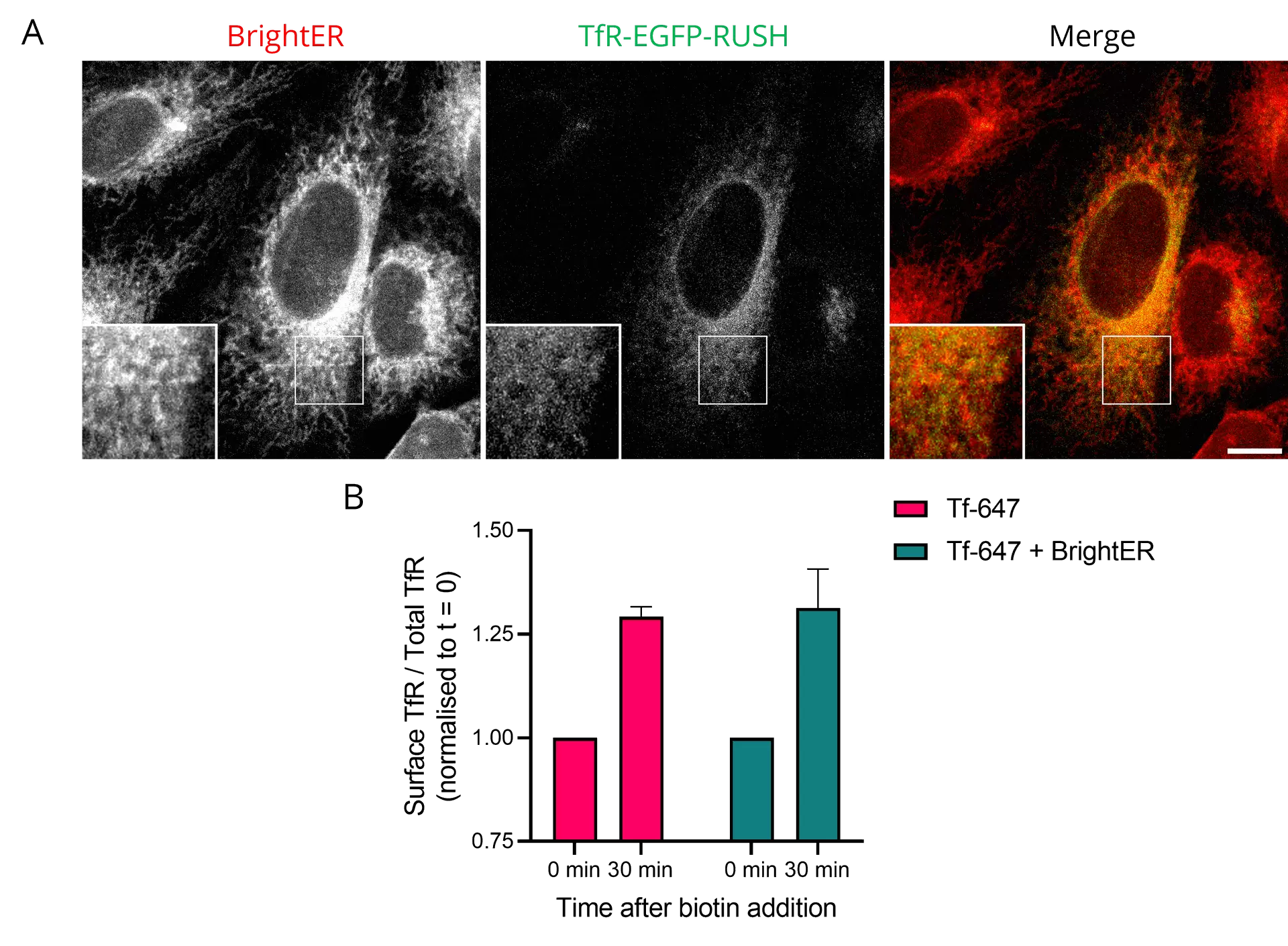
Fig.3: BrightER does not affect intracellular protein trafficking in breast cancer cells. A. The strong co-localisation of BrightER and Transferrin Receptor (TfR) fluorescence signals shows ER localisation of TfR, before biotin addition, in SUM159 cells CRISPR-edited to express the TfR-eRUSH system*. B. BrightER labeling does not alter biotin-induced TfR trafficking from the ER to the plasma membrane.
*link: https://www.science.org/doi/pdf/10.1126/sciadv.aba7803
Image credit: Raphaël Gaudin & Yonis Bare – IRIM CNRS, Université de Montpellier - 2023
- Catalog Number
RGA-RET-IDY - Supplier
Idylle - Size
- Shipping
RT