
Exosome Release & Transfer Analysis
Illuminate the complexities of EV release, transfer and subpopulation functions with EV-LuminiteTM
Illuminate the world of Extracellular Vesicles with EV-Luminite and unravel the secrets of EV releasing and transferring. Gain unparalleled insights into EV biogenesis, subpopulation EV function, and cellular communication with this cutting-edge luminous quantification tool.
- Accurate luminous quantification of EV subpopulation at remarkable low concentrations
- Sensitivity and reliability empowered by nano luciferase technology
- Easy-to-use stable Lenti-based system
- Enhanced visibility into EV subpopulation processes and functions
- Versatile applications including High Content Screening (HCS)
- Advanced research and therapeutic potential
How it Works
EV- Luminite works by utilizing nano luciferase detection system to quantify the production/secretion of Extracellular Vesicles (EVs), evaluate EV delivery and even tracing the cellular communication of EV.
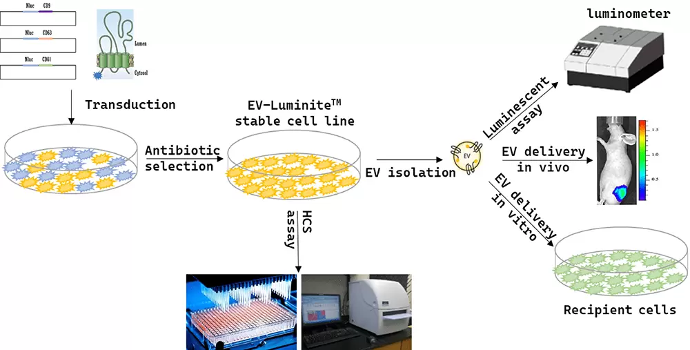
Target cells are transduced with EV-Luminite lentivirus to generate a EV-Luminite stable cell line from which EVs will be secreted.
EVs are isolated from the supernatant of the stable cell line and can be subsequently:
- Qualified by nano luciferase assay
- Delivered to recipient cells to assess EV delivery/uptaken
- Delivered in mice to study EV dynamic biodistribution
The EV-Luminite stable cell line can be used for any customized high-content screening, such as screening for compound that can increase EV secretion, et al.
EV-Luminite Constructs
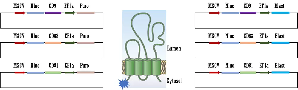
Nluc enzyme was cloned into CD9, CD63, or CD81 encoding lenti construct at the N-terminal position with either puromycin or blasticidin resistant. Middle: Scheme of the topology of a tetraspanin (CD9, CD63, or CD81) with N-terminal Nluc-tag.
EV-Luminite constructs are available as expression lentivectors, prepacked lentiviruses and EV secreting stable cell lines. In addition, as the most time-saving option, we offer purified exosomes from EV-Luminite stable cell lines.
Accurate and Sensitive Quantification of EVs

EV-Luminite allows accurate and sensitive quantification of EV. EV from Nluc-CD63 EV-Luminite stable cell line was isolated using ExoQuick-TC. A) EV samples with different protein amount were used for measuring the nano luciferase activity by Nano-Glo Luciferase Assay. B) EV samples with series dilution were used for measuring the nano luciferase activity by Nano-Glo Luciferase Assay and particle number by NTA. Nano luciferase activity shows nice correlation with EV protein amount and particle number.