Everspark 1.0 Super-resolution microscopy mounting buffer
For multicolor, long-term and stable fluorescence imaging

Product Description
Prepare your samples one day, and image them every day for weeks.
Compatible Fluorophores
Yellow to far-red. Validated fluorophores include JF 549, DL 550, CF 555, CF 568, AF 568,JF 646, AF 647, CF 647, Atto 647N, DL 650, CF 680 & Cy5
Features
Multicolor imaging: +
Blinking stability (after mounting): up to 2 months
Blinking events: +
Related Product
Everspark 2.0 Super-resolution microscopy mounting buffer with different compatible fluorophores (green, yellow & far-red) and improved imaging and blinking features.
Applications
- Use this hassle-free buffer to decouple the preparation of your samples and their imaging with dSTORM
- Prepare your samples in your lab, and image them in your core facility 3 to 4 weeks later
- Use different colors either at the same time or one after the other
How it Works
Use with a glass-bottom dish
Use with a depression slide
Supporting Data

Figure 1. Long-term imaging with Everspark 1.0. Left: Two centrosomes imaged the same Day (D0) and 7 days after mounting (D7) on the same slide stored in the dark at 4°C. The typical 450 nm donut-like structure is visualised using a colour-coded scale encoded with the IGOR software, where each point appears as a function of its localisation precision (5 to 60 nm; inverted rainbow colour scale). Labeling: Distal-appendages detected by immunofluorescence with AF647 in RPE-1 cells.
Right: The number of blinking events per centrosome and the median of the localization precision in nm are presented for each serie of 50,000 images recorded at D0 and D7 (left).
Credits: Camille Fourneaux & Karine Monier, CRCL, Lyon
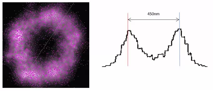
Figure 2. Donut-like structure of in-cellulo mature centrosome reconstructed after dSTORM imaging in Everspark 1.0 buffer.
Each point is represented by its centroid (purple points) and its gaussian width (white). Intensity is displayed on the right with the measurement from peak to peak, in agreement with the size of the distal appendage crone. Labeling: Distal appendages detected by immunofluorescence with AF647 in RPE-1 cells. Credit: @Corentin Rousset, Karine Monier, CRCL, Lyon
- Catalog Number
KMO-ETE-450-IDY - Supplier
Idylle - Size
- Shipping
Blue Ice