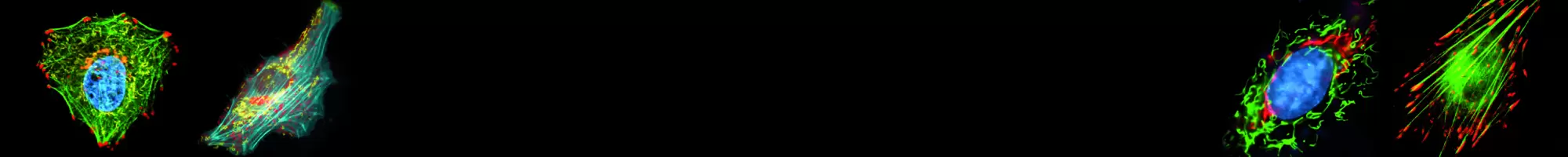
Fluorescent Proteins
Live Cell Imaging
Fluorescent proteins can be used for in vivo protein localization and interaction studies, analysis of promoter activity in live cells, tracking subcellular organelles, identification and isolation of specific populations of cells, generation of stably transfected cell lines, and more.
Ranging in color from blue to far-red, fluorescent proteins allow for the visualization of multiple events simultaneously by both fluorescent microscopy and flow cytometry. All fluorescent proteins are improved by mutagenesis and codon usage optimization for high expression levels in mammalian cells and fast maturation at 37°C.
The proteins possess bright stable fluorescence and enable monitoring of target cells or proteins over an extended period of time. No addition of cofactors or substrates is required for fluorescent protein detection.
Ready-to-use Subcellular Localization Vectors
Subcellular localization vectors express a fluorescent protein fused to a specific protein tag directing the fusion protein to the desired subcellular location.