Photoactivatable Fluorescent Protein Vectors
Photoactivatable Fluorescent Proteins
Monitoring of cellular events
Photoactivatable fluorescent proteins (PAFPs) represent a unique tool for monitoring cellular events. PAFPs change spectral properties in response to irradiation with specific light. In recent years PAFPs have been utilized for the development of novel methods for the optical labeling and tracking living cells, organelles, and intracellular molecules in a spatio-temporal manner. PAFPs became indispensible tools for super-resolution imaging techniques. Moreover, PAFPs opened new possibilities to study cell physiology, in particular, they allow careful determination of protein half-life.
Kindling Red Fluorescent Protein KFP-Red
- Reversible or irreversible photoactivation
- Activated by green light that does not damage cells and tissues
- Quenching by blue light
- Recommended for tracking cells and cellular organelle movements
KFP-Red (also referred to as KFP1) is a photoactivatable GFP-like protein generated on the basis of Anemonia sulcata chromoprotein, asFP595 [Lukyanov et al., 2000; Chudakov et al., 2003a; Chudakov et al., 2003b]. KFP-Red switches from a non-fluorescent to a red fluorescent form (with excitation/emission maxima at 580 nm and 600 nm, respectively) under the exposure to intense green light irradiation. A green light laser does not damage cells and tissues. Activated KFP-Red can be easily detected because its emission spectrum is beyond the region of cell autofluorescence.
KFP-Red can be used for in vivo monitoring cell and cellular organelle movement.
Chudakov DM, Belousov VV, Zaraisky AG, Novoselov VV, Staroverov DB, Zorov DB, Lukyanov S, Lukyanov KA. Kindling fluorescent proteins for precise in vivo photolabeling. Nat Biotechnol. 2003a; 21 (2):191-4. / pmid: 12524551
Chudakov DM, Feofanov AV, Mudrik NN, Lukyanov S, Lukyanov KA. Chromophore environment provides clue to "kindling fluorescent protein" riddle. J Biol Chem. 2003b; 278 (9):7215-9. / pmid: 12496281
Lukyanov KA, Fradkov AF, Gurskaya NG, Matz MV, Labas YA, Savitsky AP, Markelov ML, Zaraisky AG, Zhao X, Fang Y, Tan W, Lukyanov SA. Natural animal coloration can Be determined by a nonfluorescent green fluorescent protein homolog. J Biol Chem. 2000; 275 (34):25879-82. / pmid: 10852900
Photoactivatable Red Fluorescent Protein PA-TagRFP
- Monomer, successful performance in fusions
- Non-fluorescent before photoactivation
- Irreversible photoactivation to a red fluorescent form by UV-violet light irradiation
- High brightness and photostability
- Recommended for super-resolution imaging
PA-TagRFP is a photoactivatable mutant of the bright monomeric red fluorescent protein TagRFP [Subach et al., 2010]. PA-TagRFP is capable of irreversible photoconversion from non-fluorescent to red fluorescent form (with excitation/emission maxima at 562 nm and 595 nm, respectively) in response to UV-violet light irradiation.
High brightness, photostability and monomeric nature of PA-TagRFP make it an excellent protein tag for both conventional microscopy and super-resolution PALM imaging techniques [Subach et al., 2010].
Subach FV, Patterson GH, Renz M, Lippincott-Schwartz J, Verkhusha VV. Bright monomeric photoactivatable red fluorescent protein for two-color super-resolution sptPALM of live cells. J Am Chem Soc. 2010; 132 (18):6481-91. doi: 10.1021/ja100906g / pmid: 20394363
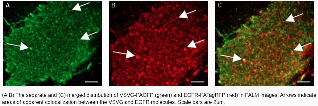
PA-TagRFP use in PALM imaging techniques
Tracking of PA-TagRFP-tagged epidermal growth factor receptor (EGFR-PATagRFP) and PAGFP-tagged vesicular stomatitus virus G protein tsO45 (VSVG-PAGFP) in live COS-7 cells by two-color single-particle tracking PALM.